


Introduction
The spleen is an intraperitoneal organ, and located in the left upper quadrant, fixed with the gastrosplenic, splenocolic, and splenorenal ligaments [1]. Wandering spleen (also known as ectopic spleen) is a condition in which the spleen is not found in its usual location or presentation. The entity is caused by absence or laxity of the ligaments fixing the spleen. Due to this laxity, splenic vascular pedicle can be twisted easily, incurring splenic torsion. Splenic torsion leads to infarction, requiring prompt diagnosis and treatment [1-3]. In this report, we present 2 cases of splenic infarction due to torsion of wandering spleen, which was successfully treated with laparoscopic splenectomy. This study was approved by the Institutional Review Board (IRB No. 05-2019-157).
Case
1. Case 1
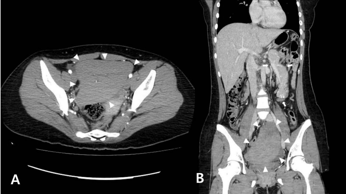
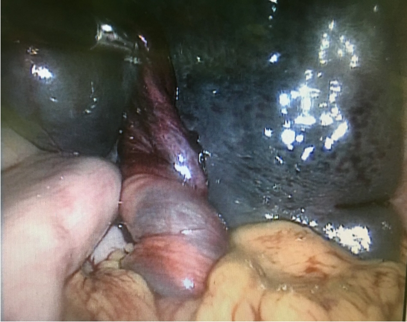
A previously healthy 17-year-old girl was referred to the emergency department due to sustained lower abdominal pain with vomiting lasting for 7 days. Prior to the referral, she had been hospitalized for 3 days at a local clinic without improvement. The initial vital signs were as follows: blood pressure, 110/75 mmHg; heart rate, 90 beats per minute; respiratory rate, 20 breaths per minute; temperature, 36.5°C. Physical examination showed localized tenderness and palpable mass at the left lower quadrant of abdomen. Laboratory examination showed a serum concentration of C-reactive protein of 1.15 mg/dL. A computed tomography (CT) scan performed at the local clinic showed a wandering spleen located at the pelvic cavity with total infarction (Fig. 1). Prompt laparoscopic exploration revealed a splenic infarction (Fig. 2), and laparoscopic total splenectomy was performed. She was discharged on postoperative day 4 without complications. To prevent an overwhelming postsplenectomy infection syndrome, she was treated with oral antibiotics for 2 weeks and vaccinated.
2. Case 2
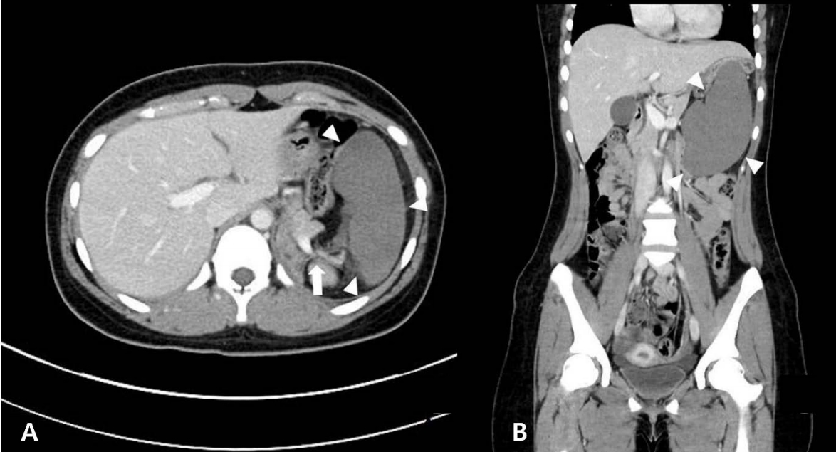
A previously healthy 15-year-old girl visited the emergency department due to severe persistent abdominal pain lasting for 1 day. The initial vital signs were as follows: blood pressure, 120/80 mmHg; heart rate, 95 beats per minute; respiratory rate, 20 breaths per minute; temperature, 36.7°C. The left upper quadrant, palpable mass and tenderness was found. Laboratory examination showed a white blood cell count of 14,280/mm3 (segmented neutrophils, 83.0%). CT scan showed whirling sign of the splenic vessels in the poorly enhanced spleen, suggesting a splenic infarction (Fig. 3). On laparoscopic exploration, the wandering spleen leading to torsion was found at the left mid-abdomen. Laparoscopic total splenectomy was conducted. She was discharged uneventfully on postoperative day 5. Prophylactic antibiotics and vaccinations were also given.
Discussion
Wandering spleen is a rare disease and accounts for 0.1%-0.2% of splenectomy cases [4]. Its incidence has not been clearly documented because of the difficulty in recognizing nonspecific symptoms [5]. Wandering spleen is most common at ages between 20 and 40 years, and is more common in women [2]. In particular, its incidence is about 20 times higher in reproductive women than men [2]. In children, reported youngest age is 3 years [4,6], and sexual difference of incidence is reported as similar or female predominant [3,4,7]. Wandering spleen is commonly found in the lower abdomen and pelvis due to gravity, but it may remain in the left upper quadrant and simply have an abnormal rotation [1,8].
The etiology of wandering has been reported to be multifactorial, a combination of congenital and acquired factors. A developmental failure in the peritoneal attachments is a congenital factor. During the second month of embryonic development, the dorsal mesogastrium fuses with the posterior abdominal wall. This process does not occur in a patient with wandering spleen. Thus, a laxity or absence of normal supporting ligaments makes the spleen prone to torsion due to a long, mobile mesenteric vascular pedicle [3,5,8]. Meanwhile, acquired cases have been associated with parity and hormonal changes, which cause weakness of the abdominal wall and a ligamentous laxity. Other possible factors for wandering spleen are trauma, gastric distension, and splenomegaly secondary to malaria, lymphoma, chronic myeloid leukemia or lymphosarcoma [3,5,8].
Clinical features vary from asymptomatic to acute severe abdominal pain accompanying a shock. Acute abdominal pain is the most common symptom, followed by palpable abdominal mass and vomiting [4]. Cases of intestinal obstruction due to small bowel adhesion to the splenic pedicle [5] and pancreatitis due to accompanied distal pancreatic torsion [9] were also reported.
Because of variable symptoms, imaging studies are essential. Contrast-enhanced CT is the imaging study of choice [1,10]. CT scans show the spleen that is located abnormally or oriented abnormally in the left upper quadrant. Tracing the course of the splenic vessels can aid in identifying the ectopic or wandering spleen along with the whirl sign, which is specific to pedicle torsion [1]. Poorly enhanced spleen indicates an infarction [10]. Ultrasonography with Doppler is frequently used as an initial study [1,3]. Ultrasound findings suggesting splenic infraction are an increased resistive index of over 80%, a low diastolic velocity in the splenic artery, and no hilar and parenchymal blood flow [9].
In patients with torsion of wandering spleen, if the spleen is viable, splenopexy is the treatment of choice, but if infarcted or not viable, splenectomy is necessary [8]. Although watchful waiting was recommended for those with asymptomatic wandering spleen in the past, most patients recently undergo operations to prevent the torsion and infarction [1]. After the operations, prophylactic antibiotics and vaccinations against Streptococcus pneumoniae, Haemophilus influenzae, and Neisseria meningitidis should be given to prevent overwhelming postsplenectomy infection syndrome [8]. Laparoscopic surgery has the advantages in cosmetic excellence and pain relief. It is easy and safe to detorsion the spleen, confirm the viability, and perform splenopexy or splenectomy with laparoscopy [9].
In summary, splenic infarction due to torsion of wandering spleen could present with acute abdominal pain. When patients have sudden onset of severe left abdominal pain without any relevant past history, wandering spleen should be considered as a differential diagnosis. If suspected, CT scan should be performed quickly, and subsequent prompt surgical interventions, splenopexy or splenectomy, are needed. In this process, laparoscopic approach should be considered.